Electron beams are our passion and the technological foundation of our company.
In the following article we recognize the achievements of the pioneers who have contributed to the use of electron beams in science and industry.
cathode ray tube
The existence of cathode rays was discovered in 1868 by Johann Wilhelm Hittorf (1824-1914) discovered at the University of Münster during experiments with gas discharge tubes. Constructed according to Hittorf's model Ferdinand Braun (1850-1918) built his first cathode ray tube at the Eberhard Karls University of Tübingen in 1889.
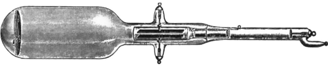
Braun's first version essentially consisted of a vacuum tube with a cathode, an anode and a fluorescent screen. By applying a high voltage, he generated a cathode ray, which he was able to detect on the fluorescent screen. In 1895 Braun was appointed director of the Physical Institute at the Kaiser Wilhelm University in Strasbourg. There he managed to focus the cathode ray with a pinhole and precisely deflect it horizontally and vertically with the help of magnetic coils. This concept became known as the Braun tube and remains the basis for controlling electron beams today.
Cathode ray tube by Ferdinand Braun (1897)
Discovery of X-rays
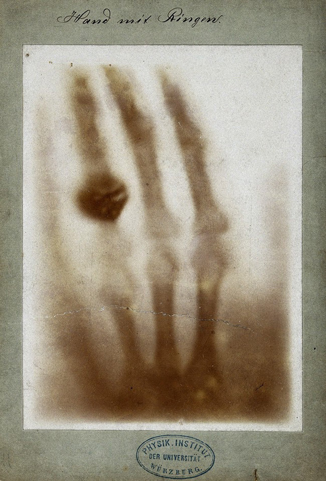
The Braun tube fascinated many scientists, including Wilhelm Conrad Röntgen (1845-1923) from the Physical Institute of the University of Würzburg. In 1895 he discovered a previously unknown type of radiation that caused luminescent paper to glow at some distance from the cathode ray tube. The X-radiation he named was created by the interaction of the cathode ray with the metallic shadow cross of the Hittorf tube. The generation of X-rays by bombarding a metallic target is still the predominant principle of technical X-ray sources today.
In 1895, the year X-radiation was discovered, Röntgen managed to take the first photographs on photographic plates. The term X-rays only became established later in German and Slavic-speaking countries.
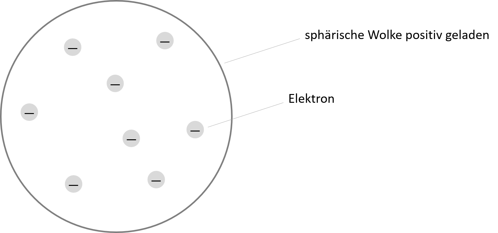
The physical nature of cathode radiation remained unclear for almost 30 years after its discovery. In 1897 the physicist experimented Joseph John Thomson (1856-1940) at the University of Cambridge with cathode ray tubes and realized that radiation consisted of small charged particles, which he called corpuscles and which later became known as electrons. Thomson is considered the discoverer of the electron and a pioneer of the modern atomic model.
Atomic model according to Joseph John Thomson (1897)
Advances in electron production
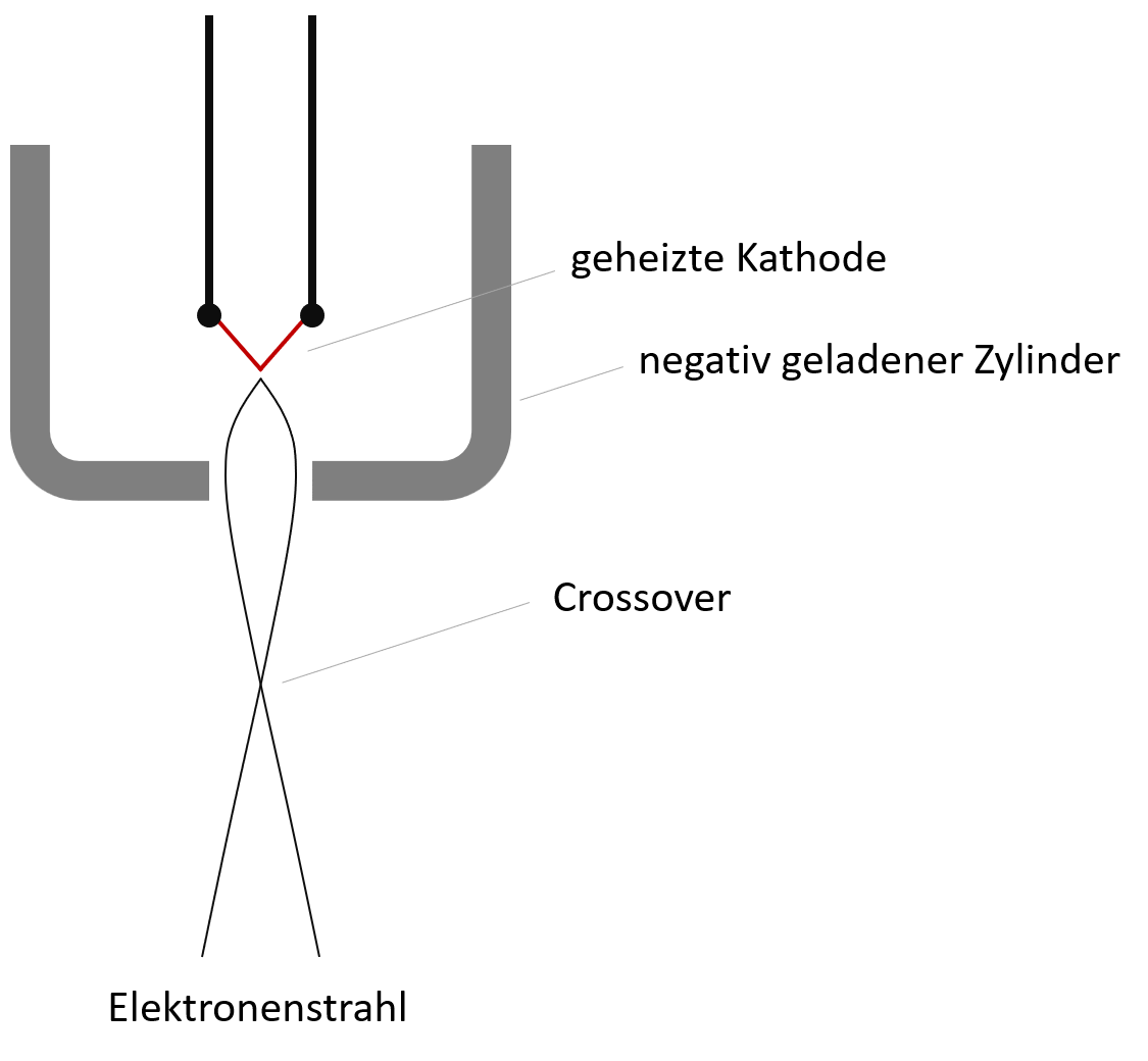
In 1902 it succeeded Arthur Wehnelt (1871-1944), who taught as a professor at the Friedrich-Alexander University of Erlangen, significantly improved electron generation by developing a heated cathode. Thermal emission enabled the extraction of electrons at significantly lower anode voltages.
At the same time, Wehnelt proposed the use of a negatively charged cylinder enclosing the cathode. The Wehnelt cylinder causes the beam to be focused and allows the brightness to be controlled by varying the applied potential. Wehnelt's work still forms the basis for the technical production of electron beams and X-ray sources today.
Improved electron generation according to Wehnelt (1905)
Resolution and wavelength
Already calculated in 1873 Ernst Abbe at the University of Jena the resolution limit of microscopes and realized that this lies in the wavelength range of the light used. This principle limit is known as the Abbe limit in optics.
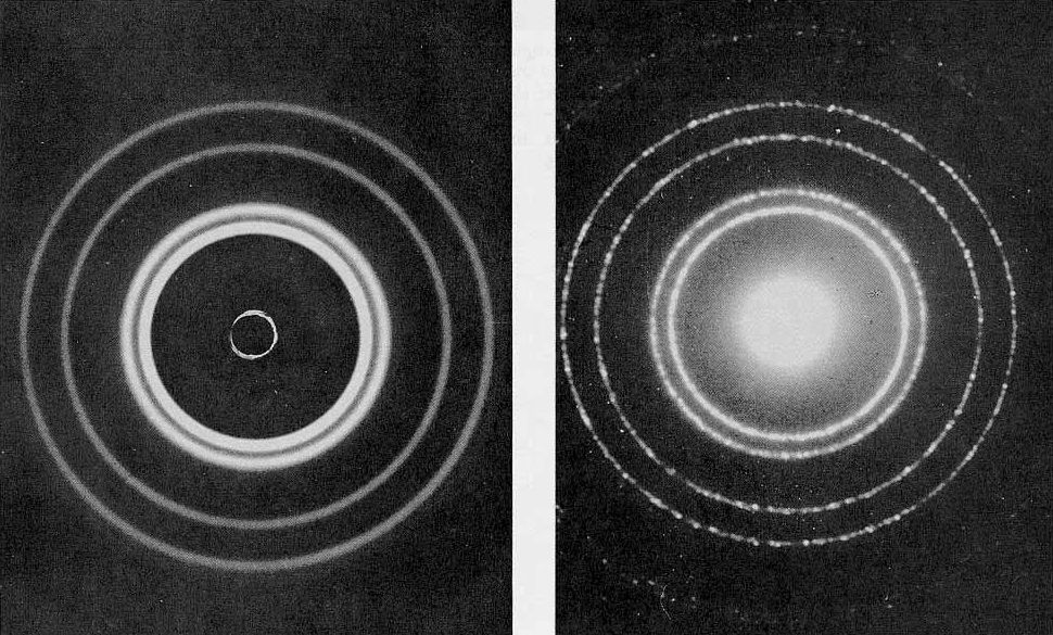
Postulated in 1924 Louis de Broglie (1892-1987) that particles with mass also have a wave character. For electrons undergoing an acceleration of 15 kV, he calculated a wavelength of less than one nanometer. The experimental proof of his hypothesis was successful Davisson and Germer in 1927 by diffraction of an electron beam on a nickel crystal.
Electron diffraction pattern of Davisson-Germer (Al crystal)
Electron microscopy
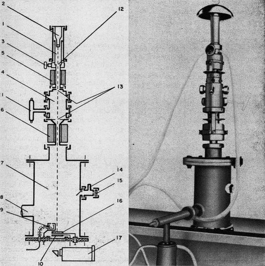
Ernst Ruska (1906-1988) and Max Knoll (1897-1969) recognized the revolutionary potential of the extremely short de Broglie wavelength of electrons to shift the Abbe limit of microscopy down to the nanometer range.
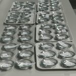
Ruska worked for Knoll at the Technical University of Berlin and built the world's first electron microscope as a doctoral student in 1931. He constructed the electron source based on Wehnelt's model with thermal emission and electrostatic focusing. Ruska succeeded in developing a second-stage magnetic lens that achieved 12,000x magnification. His electron microscope worked in transmission mode (TEM), and he projected the electron image onto a ground glass. To test magnification, Ruska used small metal grids.
Another breakthrough was achieved Manfred of Ardenne (1907-1997), who developed scanning scanning (SEM) for electron microscopy in 1937. In this mode, the object to be examined is scanned point by point with the electron beam. Von Ardenne used Braun tube technology to control the beam. Scanning operation paved the way for numerous new detection principles in imaging and spectroscopy, including microscale elemental analysis (EDX).
Transmission electron microscope by Ernst Ruska (1931)
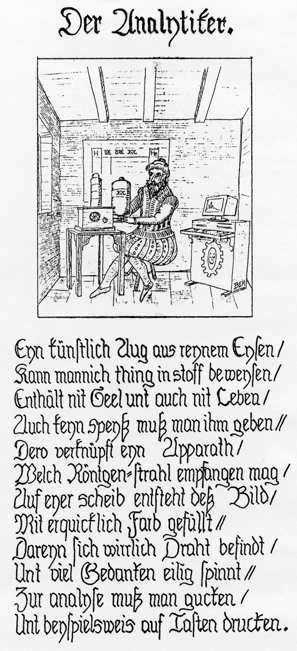
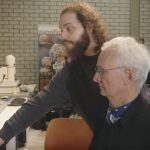
Our company founder Bernhard E. Heneka was an enthusiastic expert in X-ray element analysis in the micro range (EDX) but also an amateur painter. Bernhard E. Heneka began to explore his vocation artistically as early as the 1980s.