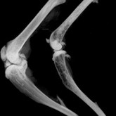
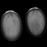
In breast cancer therapy, the precise detection of microcalcifications and clips in the specimen is essential. Within just a few years, 3D tomosynthesis has established itself as the gold standard for margin assessment in the USA.
The latest detector technology made of amorphous selenium (a-Se) delivers even higher image contrast with the shortest shutter speeds. Kubtec has used the enormous advantages of a-Se in the new PICASSO series. Intraoperative margin assessment and pathology in particular benefit from this.
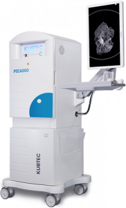
Kubtec develops innovative X-ray technologies under the trade name Kubtec Medical and ScientificThe cabinets are fully shielded and correspond to full protection devices, so the systems can be used anywhere.
Embedded microcalcifications are typical of ductal in situ carcinomas. In breast cancer treatment, the marked tissue is removed during surgery. Kubtec's compact X-ray machines allow for rapid assessment of the resection margin, as the microcalcifications are clearly visible on the X-ray image. The Kubtec systems are mobile and can be used directly in the operating room if necessary. This saves valuable time!
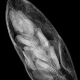
The use of specimen radiography also leads to considerable time savings in pathology. The clip can be found quickly in the X-ray image, so that the samples can be prepared precisely and with just a few cuts.
unique selling point
Optical Imaging & Image Blender
The XPERT, MOZART and PICASSO radiography systems are equipped with an optical HD camera. A high-resolution color image is automatically recorded along with the X-ray image. The Image Blender can be used to superimpose the two images. The function is patented and can only be offered by Kubtec.
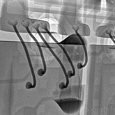
Quickly find the clip in preparations
Photographic documentation of the preparations
DIGIMUS software
DXA analysis in small rodents in vivo
Dual X-ray absorptiometry (DXA) enables the quantitative determination of body composition through image analysis at two X-ray energies. Typical measurements are the proportions of bone, fat and soft tissue as well as bone density (BMD). The DIGIMUS software controls the X-ray device for the DXA scan and guides the user through the image analysis to the result.
Digital X-ray imaging is increasingly being used for the analysis of seeds. Image analysis allows seeds to be examined individually in large ensembles. Quality control is a priority for seed producers and banks. Automatic image analysis of seeds can determine germination and morphology. In addition, diseases and pest infestations can be quickly identified. In agricultural science, tracking embryo growth is an important topic that is increasingly being investigated using X-rays.
Digital radiography devices are used in a variety of ways in science (e.g. biology, preclinical research) as well as in industry for non-destructive testing (e.g. medical devices, electronics). Kubtec's systems are used extensively as specimen X-ray devices in mammography (screening of punches), breast cancer therapy (assessment of incision margins) and pathology.
If you continue browsing this website, you agree to our use of cookies Data Protection to.