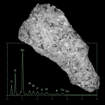
Microscopic examination & elemental analysis in one step
SEM-EDX
Functional Principle in Focus
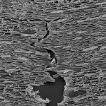
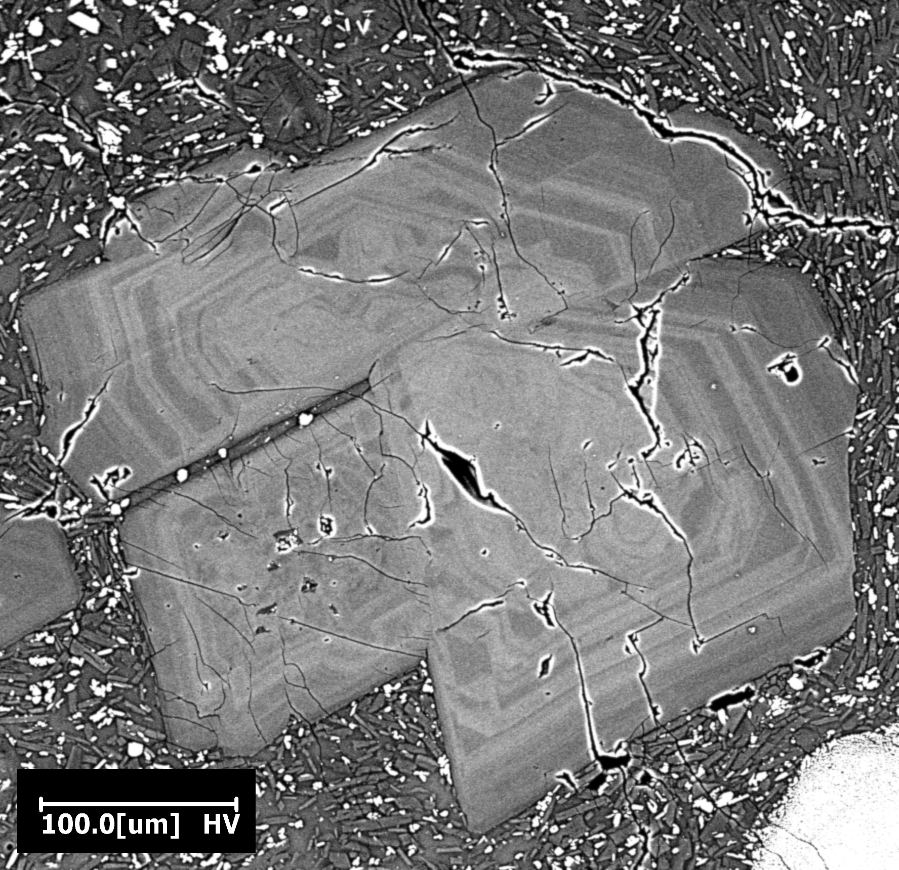
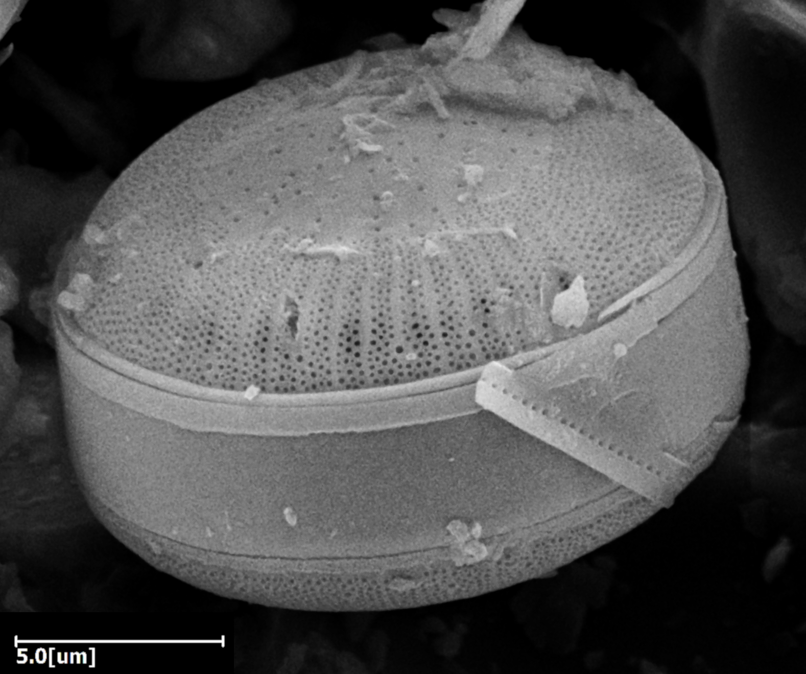
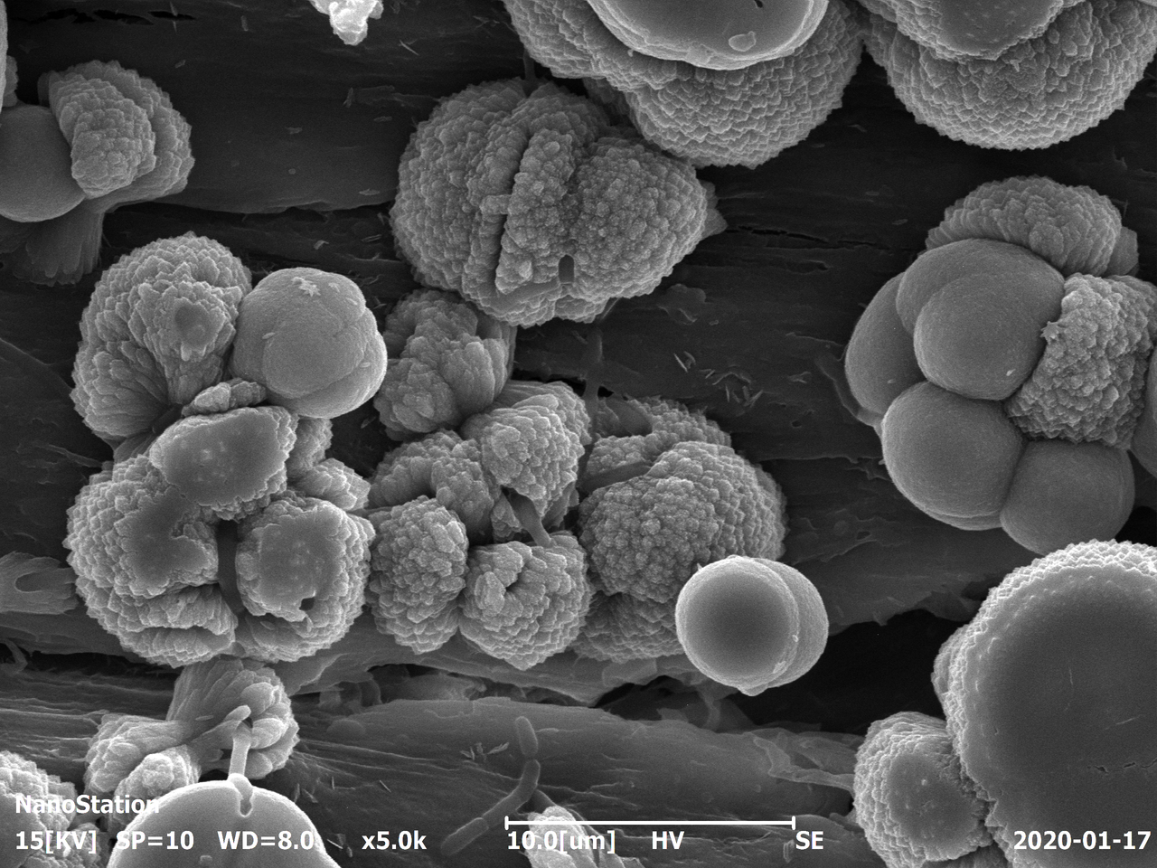
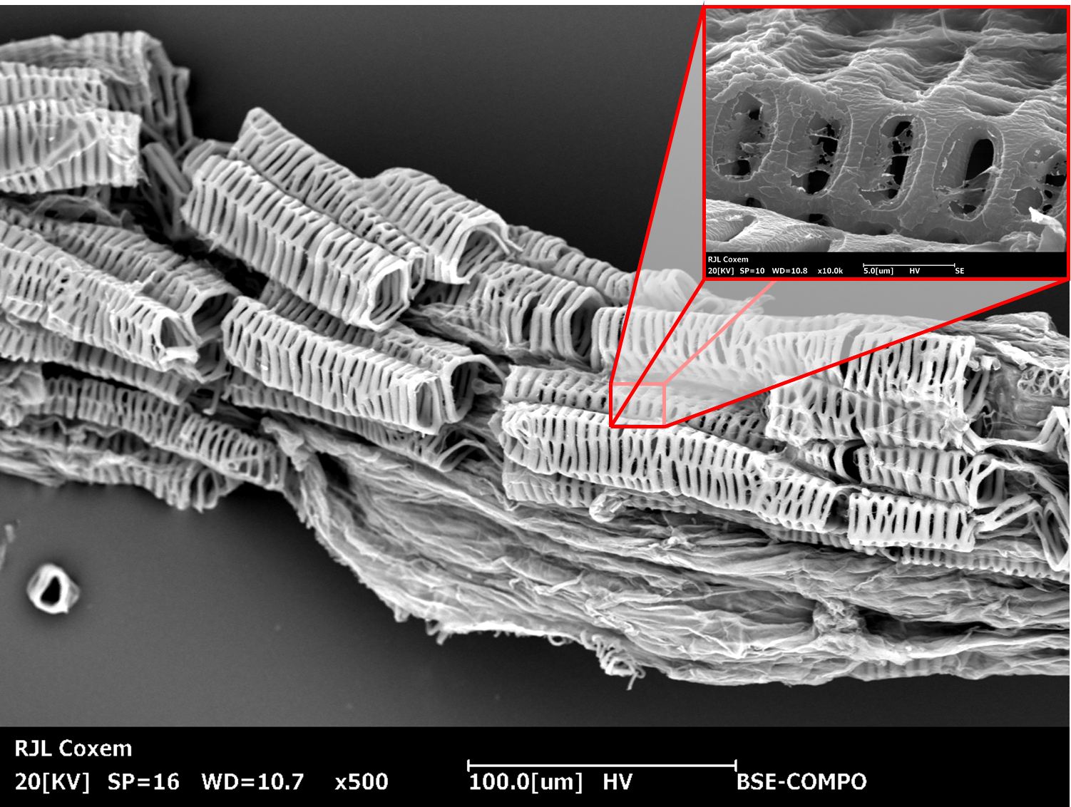
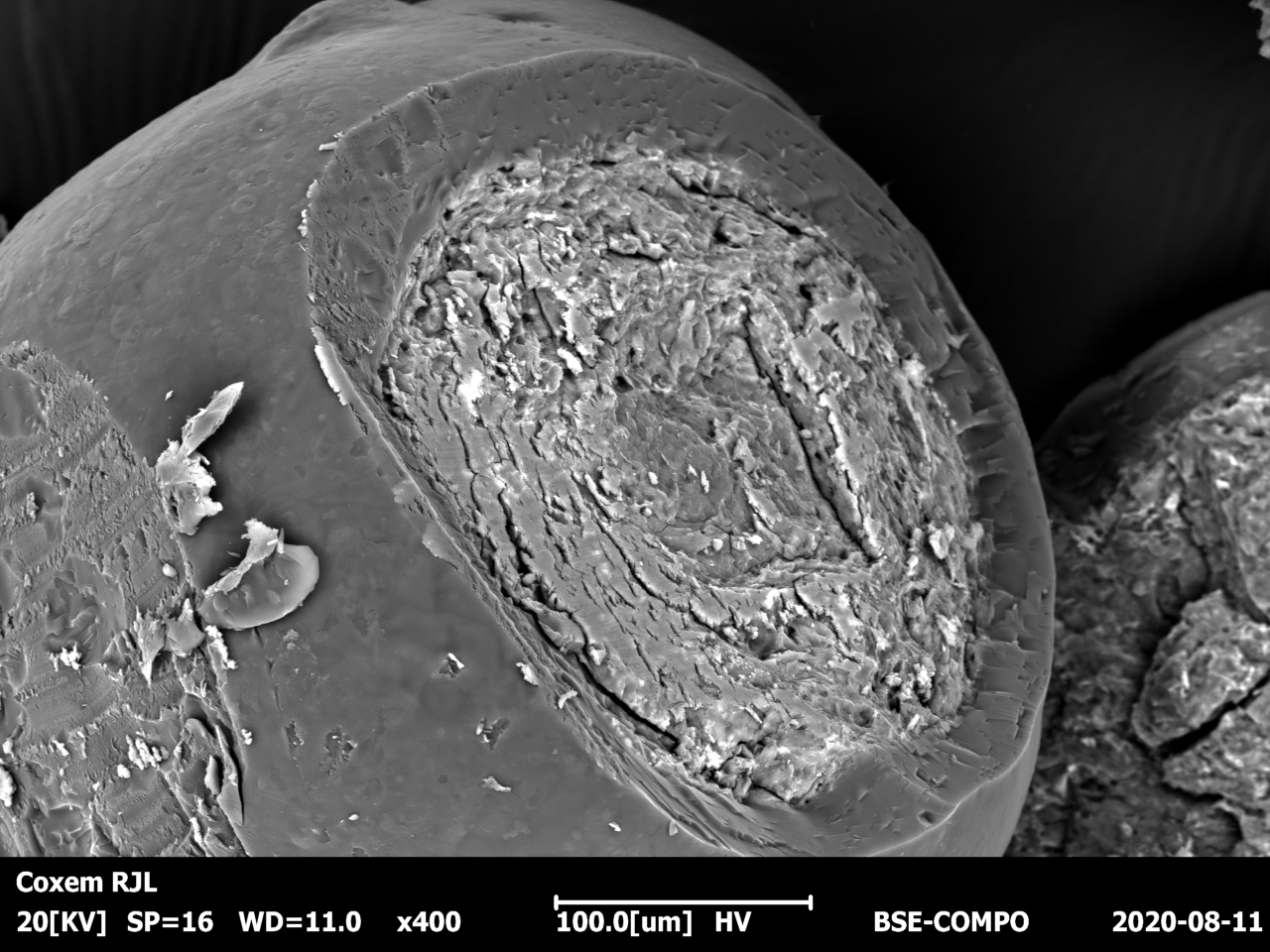
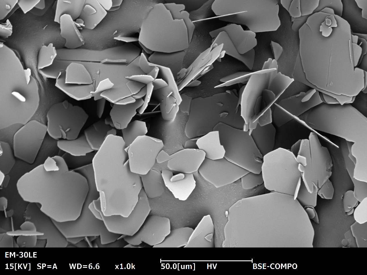
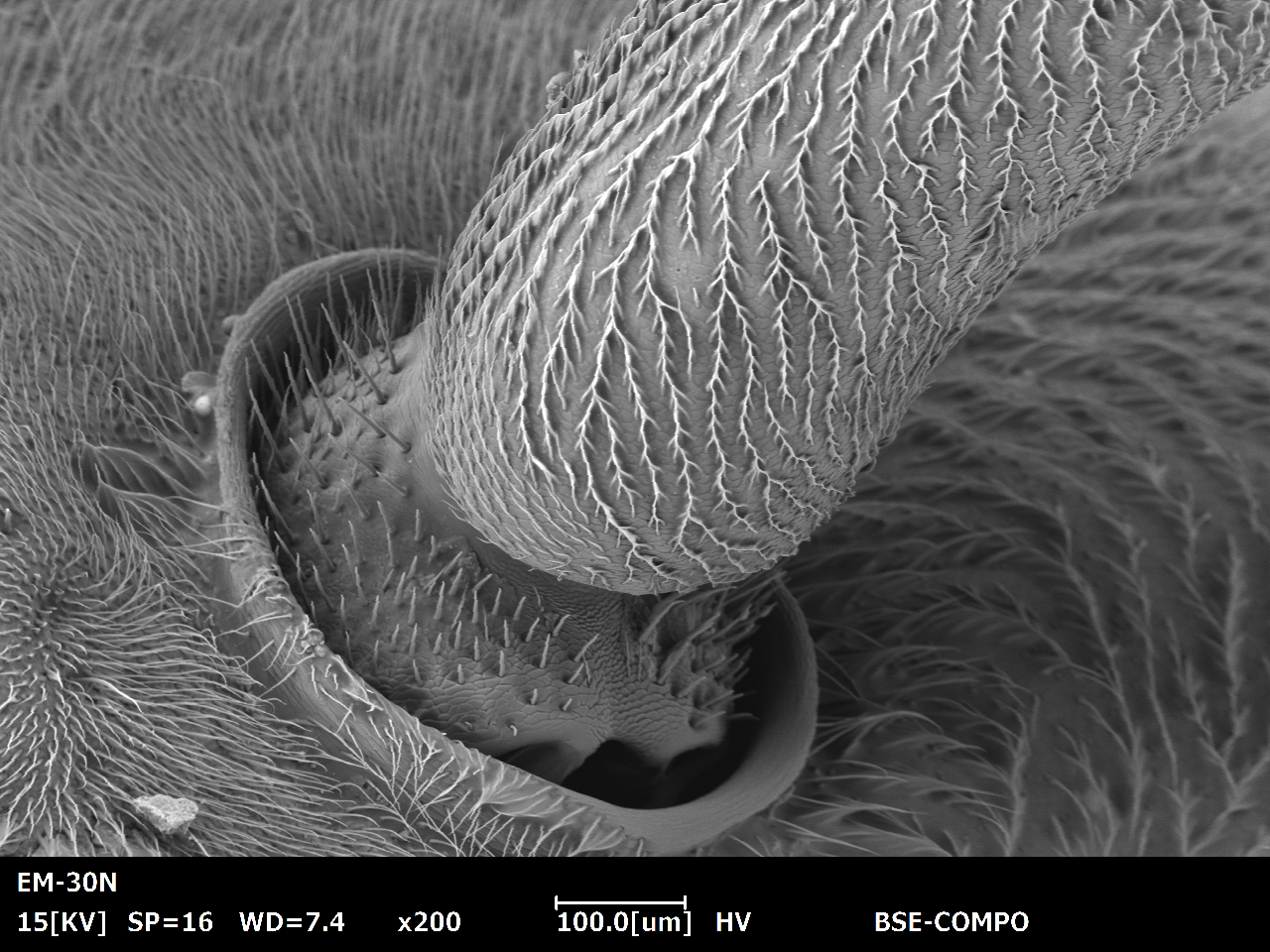
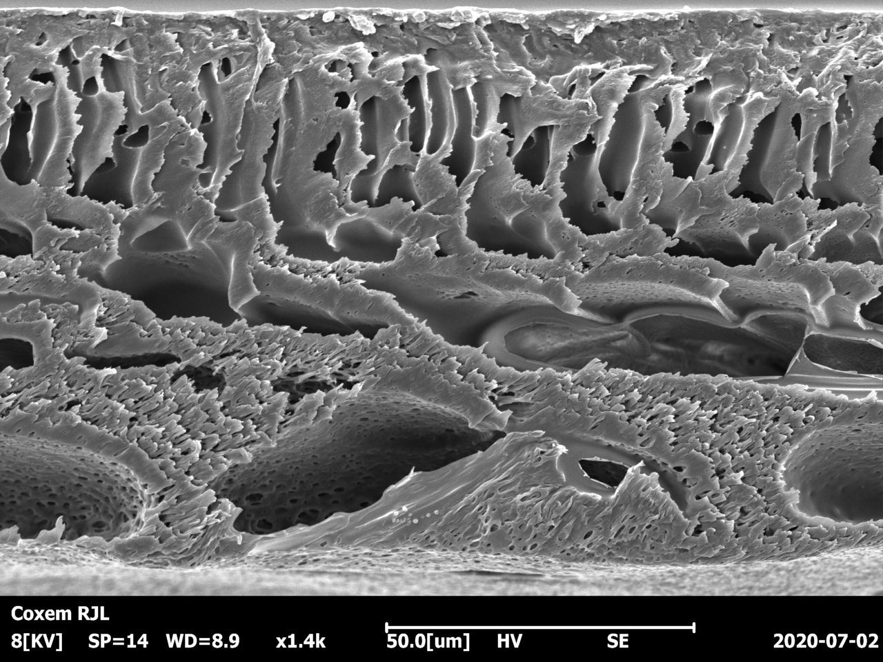
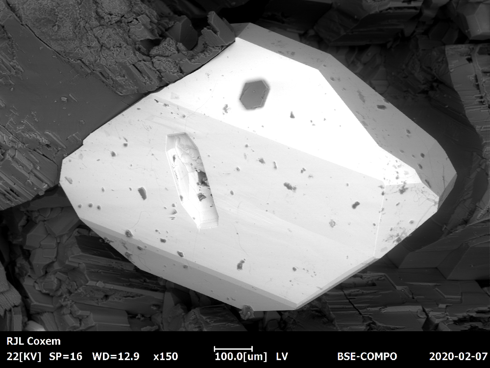
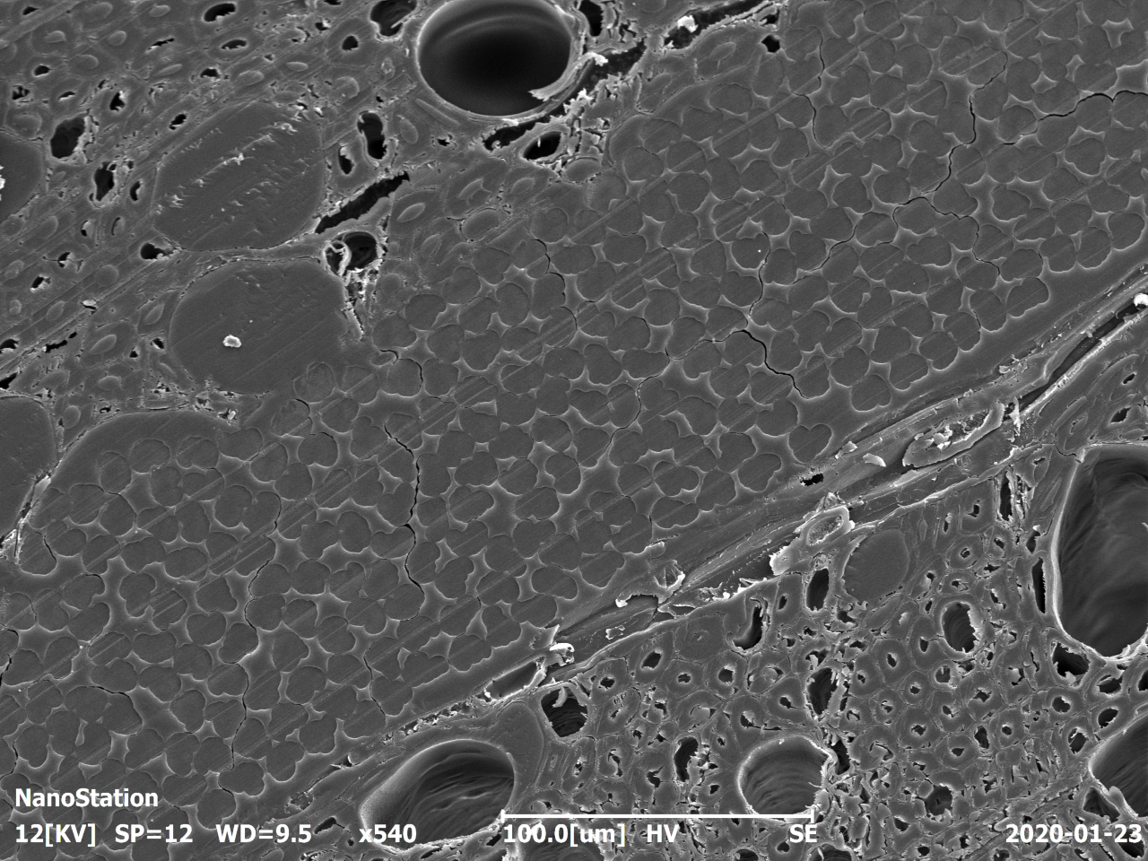
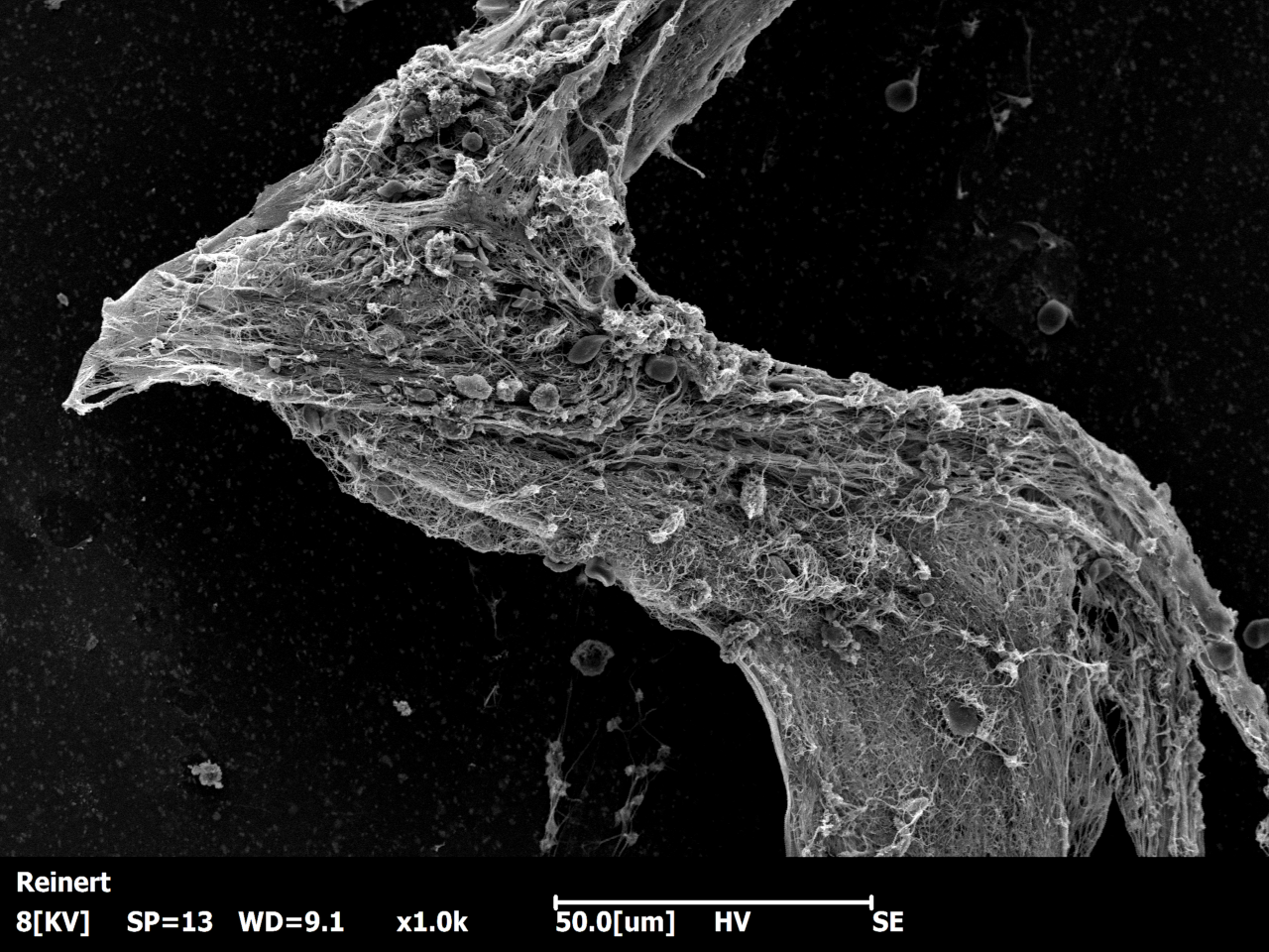
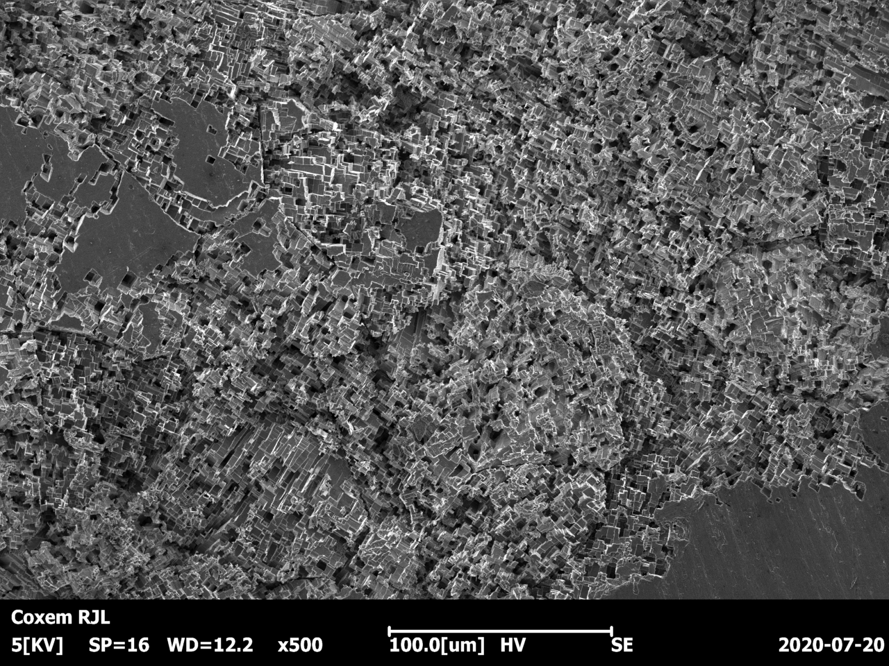
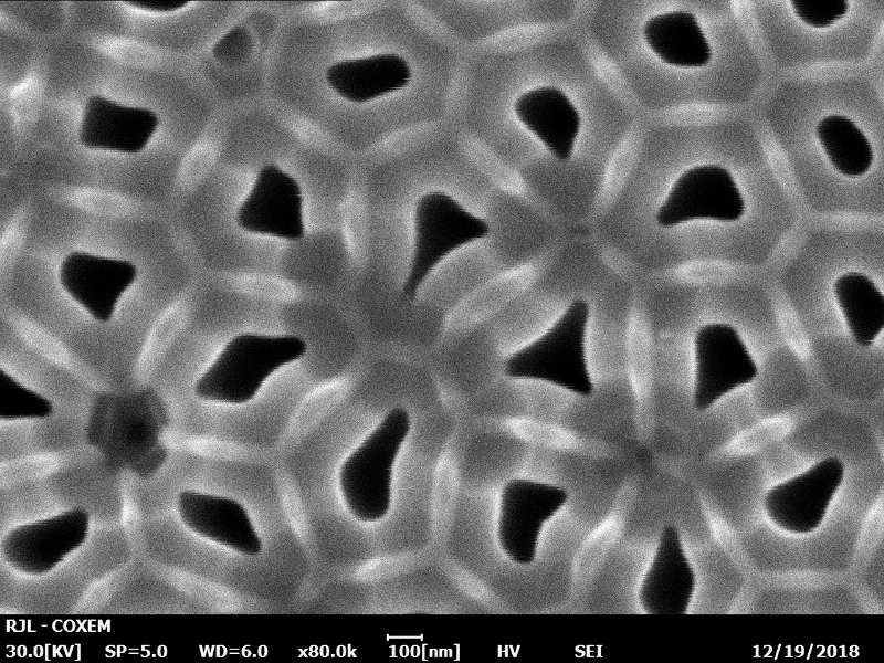
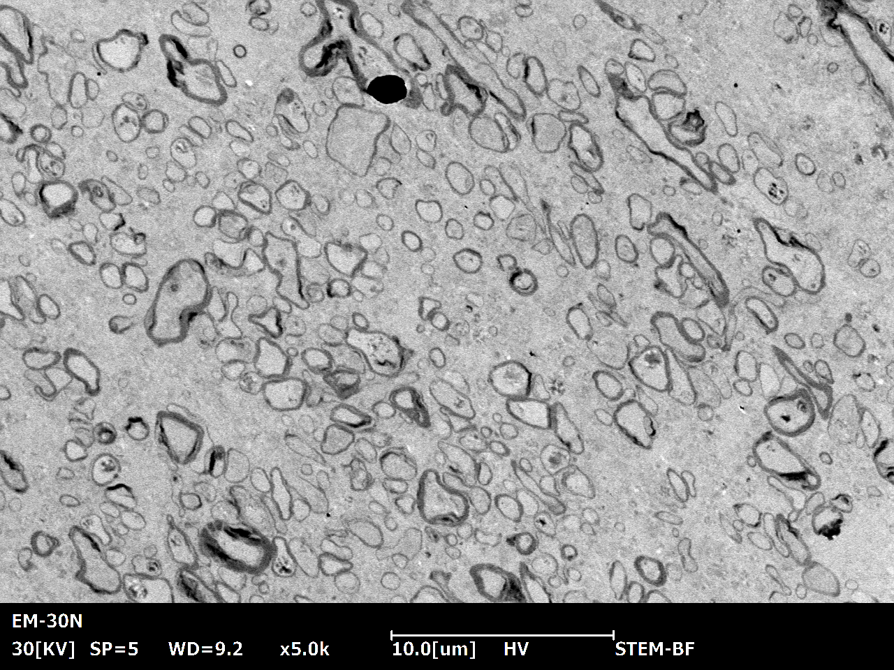
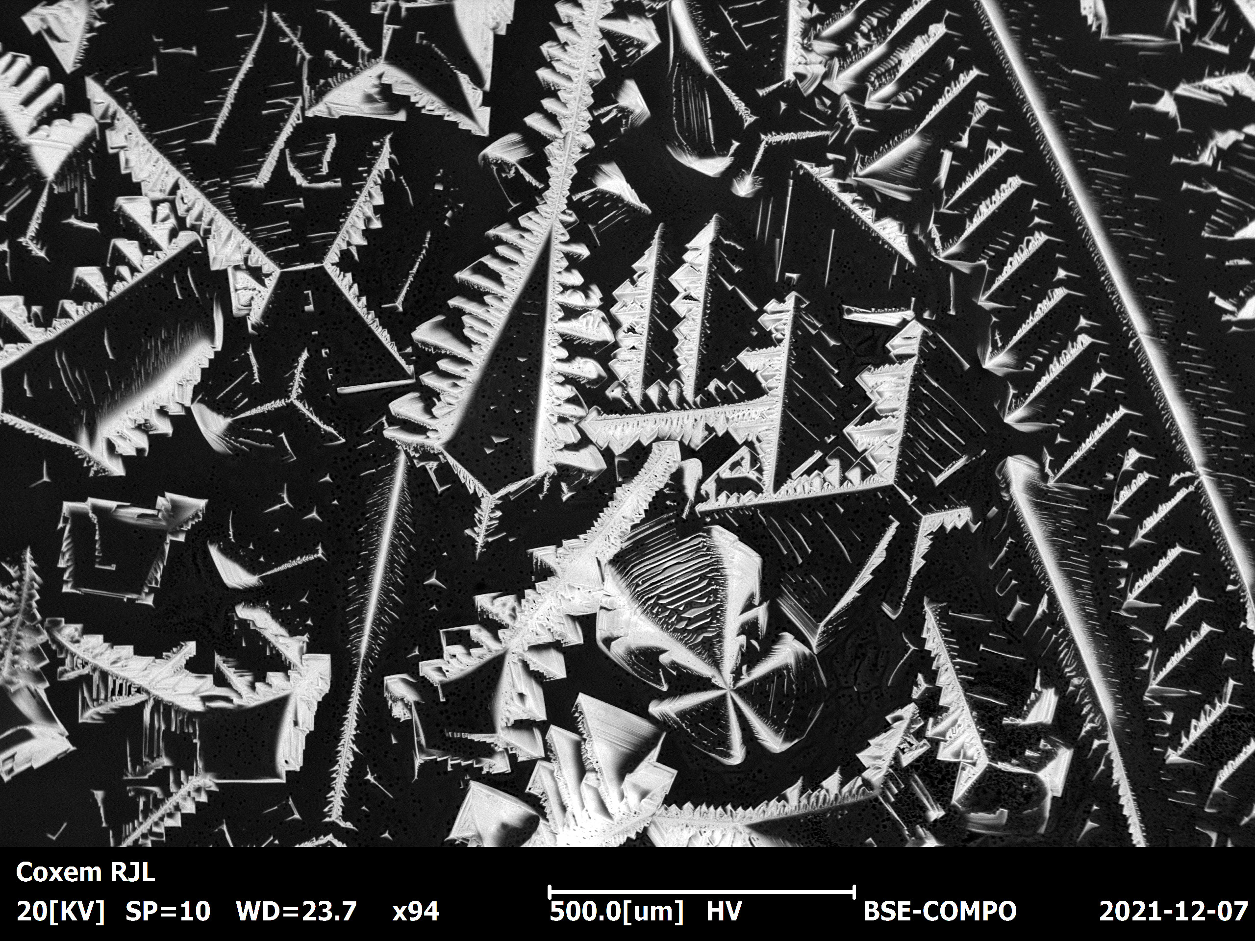
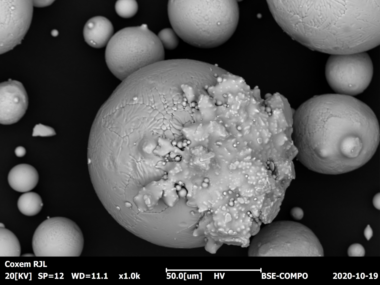
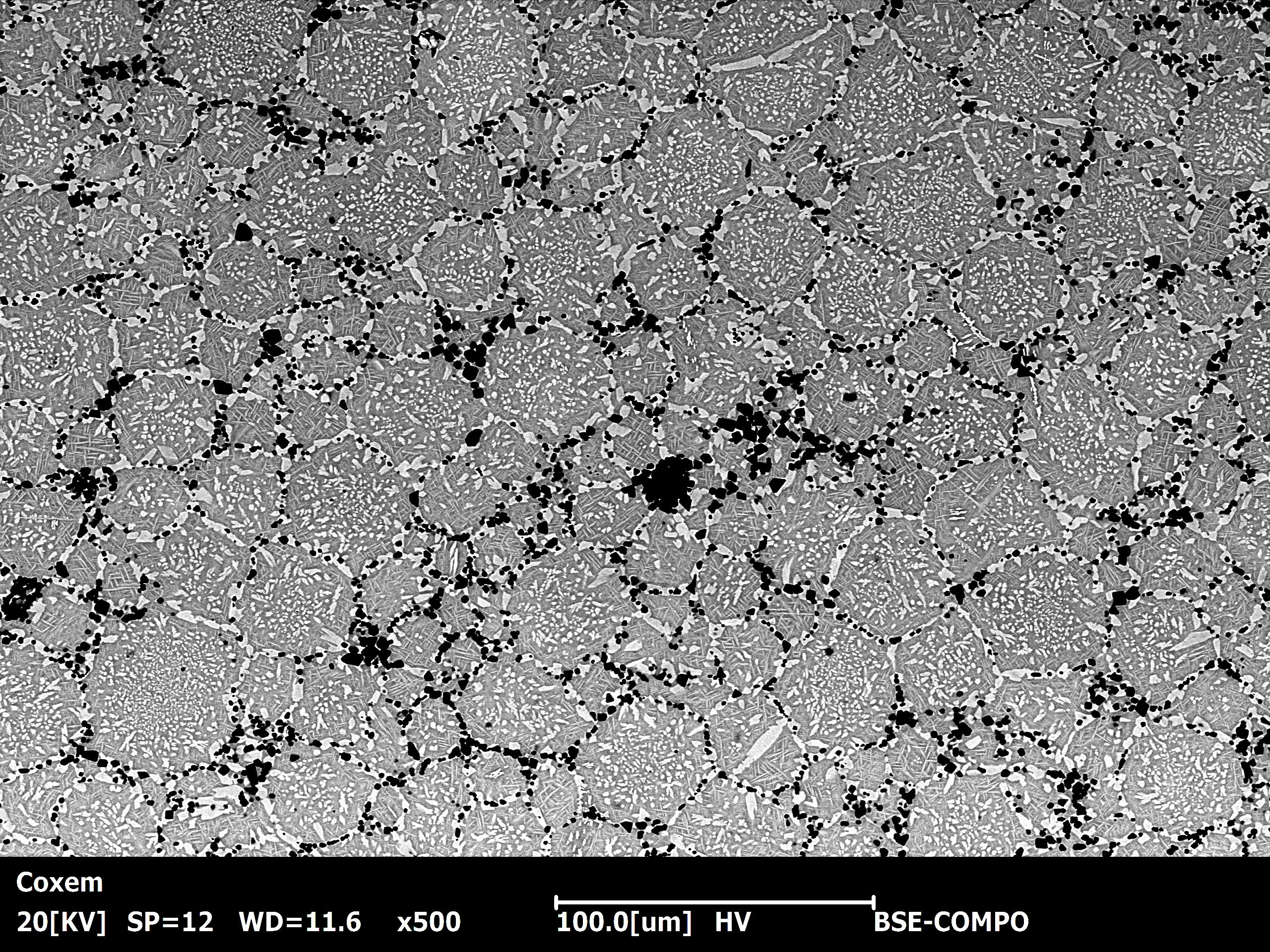
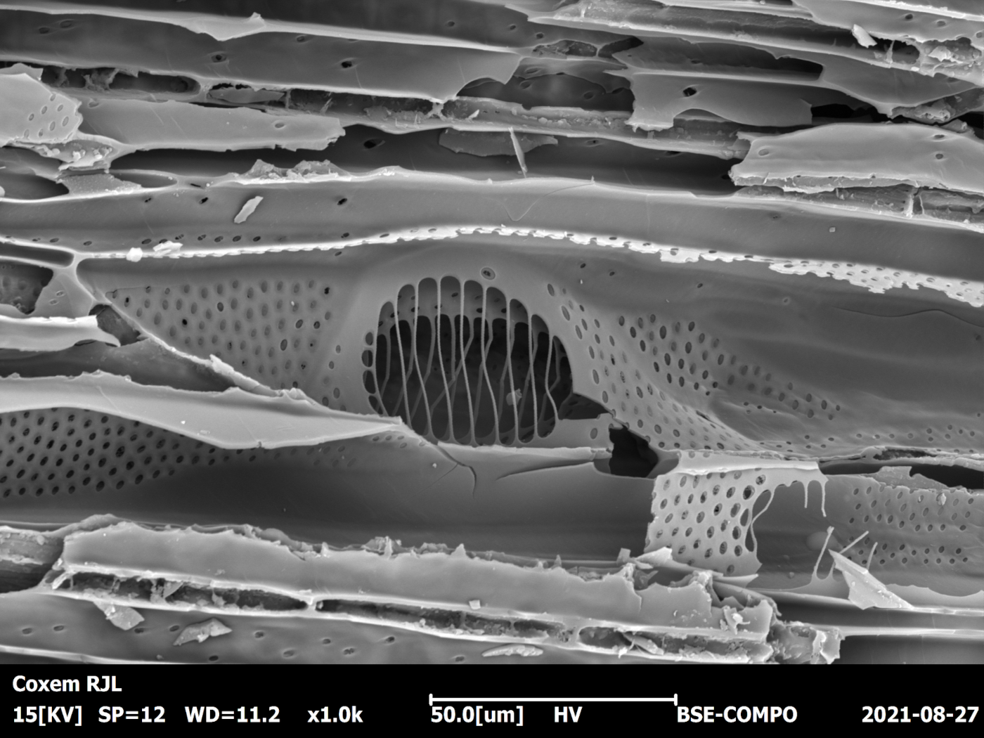
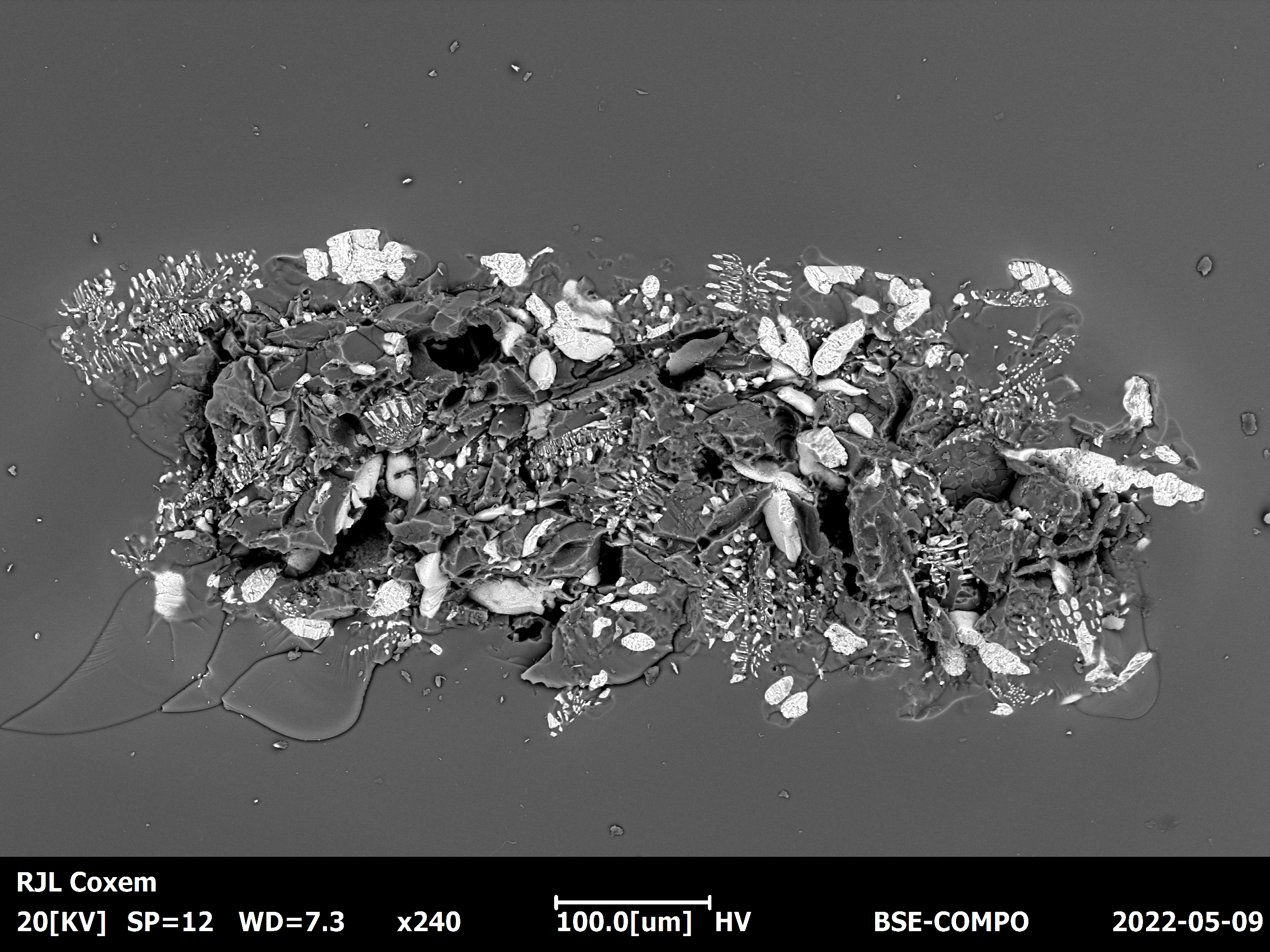
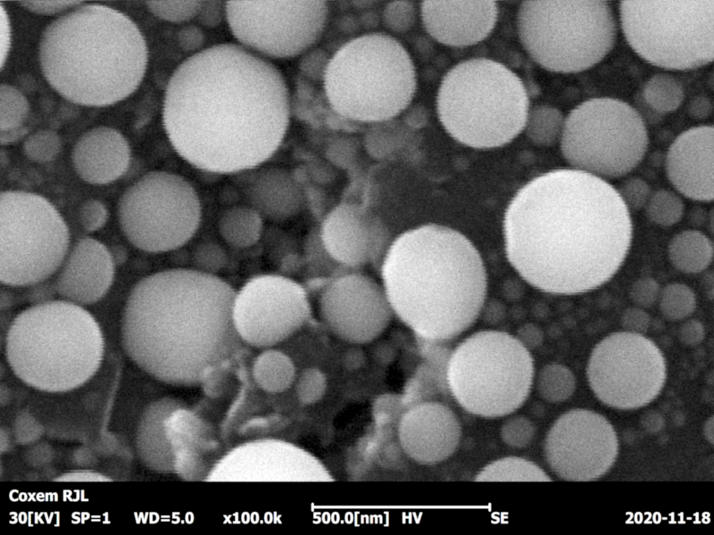
In scanning electron microscopy (SEM), a focused primary electron beam is rastered stepwise across the sample. At each pixel, the reflected electrons are detected, yielding a microscopic greyscale image of the sample. There are two imaging techniques:
Backscattered electrons (BE) → material contrast
Secondary electrons (SE) → topography contrast
In addition, the primary electron beam triggers the sample to emit characteristic X-Rays. The elements in the sample as well as their respective modal weights can be precisely determined by analyzing the color spectrum in an EDX detector.


