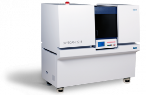


A year and a half ago we installed a Coxem EM-30N at the Eberswalde University for Sustainable Development (HNEE). The SEM-EDX tabletop device is used for examining wood-based materials. First results were published already in the same year in scientific journals.
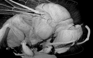
One of the users is changing his job and as a little farewell gift he made us a panorama SEM image that he took with the Coxem EM-30N. This mosaic shows a 3-point bending fracture in a deformed wood sample. With the standard integrated panorama module from Coxem, 169 individual images of a microtome section of the fracture site were collected at 200x magnification with the secondary electron detector and automatically stitched. The imaging procedure took almost 3 hours. We were particularly impressed by the stability of the electron beam during the entire acquisition time.

The compression of the fibers and the indentation at the point where the force was introduced can be clearly seen in the upper image segment. This compression was enhanced throughout the duration of experiment and the layers of wood underneath break (compress). This initial damage pattern is a so-called pressure fracture, which can only be inferred with difficulty from the outside. Further down, the stabilizing effect of the transverse vessel structures decreases more and more until the tensile strength of the wood is exceeded and a tensile fracture causes the specimen to fail completely. What is particularly interesting about this panoramic image is that the two fracture types meet. The "neutral axis" known from mechanical theory probably lies in this plane.
")
If you would like to carry out similar analyses or are interested in your own SEM-EDX system, we look forward to hearing from you.
More information about the method can be found on our SEM-EDX overview page or in our SEM-EDX guideline.