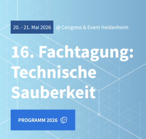
Satisfied Coxem Customer Sends a Panoramic Greeting Card

A year and a half ago we installed a Coxem EM-30N at the Eberswalde University for Sustainable Development (HNEE). This mosaic shows a 3-point bending fracture in a deformed wood sample. With the standard integrated panorama module from Coxem, 169 individual images of a microtome section of the fracture site were collected at 200x magnification with the secondary electron detector and automatically stitched.

EBSD Implemented for the First Time in a Tabletop SEM-EDX

We supported Bruker Nano in the world's first commissioning of a compact EBSD system on our Coxem Tabletop SEM-EDX. The installation went smoothly and after one hour we were able to record the first EBSD mappings ‒ Important step for the market introduction of the EBSD technology for SEM-EDX tabletop devices.

Wachstumszonen im Feldspat (BE)

Coxem Introduces BE Detector with Excellent Z Resolution

Coxem recently presented a new backscattered electron detector (BE) for their SEM-EDX systems, which impresses with its excellent sensitivity. The new BE detector is particularly interesting for applications that benefit from a high contrast of the atomic number Z.

We Support Helios in the Refurbishment of FFP Protective Masks

Given the continuing shortage of protection materials for hospital staff during the Corona crisis, Helios experts have in the past weeks developed a safe procedure for refurbishing FFP masks. Helios is making this method publically available, so that all medical staff can be supplied with sufficient protective materials.

Diatomee-Mikrofossil (SE)

Surprising Discovery of Diatoms Using SEM-EDX

Researchers from Bremen provided us with this interesting sample of volcanic ash, which we analyzed with our Coxem Tabletop SEM-EDX. In addition to the spongy structure of the fine ash particles typical of volcanic eruptions, we identified various microfossils, so-called diatoms.

Bakterielle Kalkablagerungen auf Holz (SE)

SEM-EDX Analysis of Bacterial Carbonate Deposits on Wood

Employees from the wood engineering department (Abteilung Holzingenieurwesen) sent us an aged wood sample with a typical discoloration. Using our Coxem scanning electron microscope, we imaged the microstructure in high resolution and performed an elemental analysis of the discoloration.

Tracking down the Killer Particle

When it comes to component cleanliness, the hardest particles cause the most damage. The difficulty, however, is finding these killer particles because they can be so small that they cannot be seen with a light microscope.