Application Examples of X-Ray Micro- and Nanotomography
Biological Preparations
In recent years, microtomography has proven itself as a method for the morphological characterization of animal and plant samples in zoology and botany. The advantage over histology is obvious: rare specimens can be examined non-destructively in microscopic resolution and are thus preserved for later studies.
Example: Insects & Small Creatures
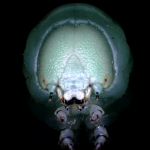
In the following example, we examined the head of a mayfly nymph (genus Coloburiscus) and visualized its masticatory muscles with an internal cross-section. We chose a high resolution of 1 μm / voxel in order to visualize the fine structures. Specimen provided by a German natural history museum.
Archaeological objects
Archeologists have long been relying on micro-CT scanners due to the benefits of a non-destructive analysis. Scientists scan tools, potsherds, funerary goods or teeth, and can also detect bone deformities, injuries and illnesses. Wood species can be assigned to charcoaled timber, and carbonized scrolls can be virtually unrolled using micro-CT.
Example: Human Remains
The following video shows the scan of the skull of an adult male individual from Thuringia, dated to the late Middle Ages, ca. 13th century. Sample provided by the University of Jena.
Preclinical Research
Preclinical research, especially in studies of laboratory mouse osteoporosis, was the first research field utilizing microtomography. In addition to the examination of bone diseases and their healing, studies on the vascular system, the lungs, the heart and other organs have nowadays become routine analyses, all benefiting from the high spatial resolution of microtomography.
Example: Femur of a Mouse (in-vivo)
In the following example, we scanned the femur of a mouse in vivo and analyzed the trabecular portion of the bone.
Example: Vascular System of a Mouse (in-vivo)
In the following example, we stained the vascular system of a mouse with contrast media and scanned it in vivo.
Example: Titanium Implant in Pig Rib
In the following example, we scanned a titanium implant that was placed in the rib of a pig ex-vivo. The interface between implant and bone is shown without artifacts, which makes it very easy to assess osseointegration.