FILTERS, COMPOSITES & FOAMS
Application Examples of X-Ray Micro- and Nanotomography
X-Ray microtomography has become a valuable tool in the analysis of lightweight materials and filter media. The 3D image allows a direct insight into the spatial distribution of fibers and pore structures. By linking with methods of numerical simulation, the relationships between structure and physical parameters such as mechanical strength and permeability can be determined directly.
Example: Fibre-Reinforced Synthetics
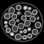
In the following example, we investigated a fiberglass-reinforced synthetic material for a manufacturer of injection-molded plastic components. These material fibers have a diameter of 5-6 μm and can be visualized with high contrast using a high-resolution scan.
Example: Foams & Lightweight Materials
The following video shows the analysis of an open-pore carbide foam made of steel. On behalf of the customer, we measured the pore size distribution and the thickness of the metal framework. In addition, the customer has used the dataset in a numerical simulation to calculate the mechanical strength and elasticity.
Example: Filter Medium
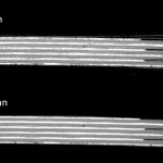
Microtomography is a suitable tool for analysing multiphase filter media by mapping the spatial structure of individual functional layers. In the following example, we scanned a two-layer filter material with a high resolution of 1 μm / voxel.


