Identification of molecules by infrared & Raman spectroscopy
Molecular Spectroscopy
Operating Principle
Infrared and Raman spectroscopy are methods of molecular spectroscopy. In both cases, the interaction of incident light with the sample molecules provides information regarding the type of molecules as well as their quantity.
During infrared spectroscopy (IR), the sample is irradiated with long wavelength light and the spectral absorption of the transmitted light examined. During Raman spectroscopy, a sample is irradiated with laser light and the wavenumber shift of the reflected light spectrally examined. In both cases, spectral peaks are indicative of a molecule type.
In our test lab, we utilize both methods of molecular spectroscopy, combined with light microscopy, Thus, we can carry out micro-scale spectroscopic analyses, e.g. on particles.
Organic fibers & dirt particles
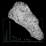
Microscopic molecular spectroscopy has proven to be very useful in the identification of unknown organic fibers and particles for the purpose of determining their origin. For this purpose, the measured spectra are compared with a reference database, and the most likely matches extracted by the software.
In the following example, we have identified an unknown dark fiber and a black particle on behalf of a customer. It has been demonstrated that the fibre is made of cotton and therefore originates from a textile. The particle, on the other hand, is made of polyphenylene sulfide (PPS), a common high-performance plastic.
Filmic Impurities
Practical example: After final cleaning, a customer repeatedly identified a filmic contamination on his components, which impeded the subsequent coating process. A spectroscopic comparison between the utilized processing auxiliaries and the unknown substance showed that the filmic contamination was a mixture of two components. The problem was solved by changing the detergent.
Chemical Imaging
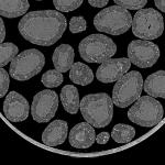
Direct chemical imaging is enabled by molecular spectroscopy. At each pixel, a spectrum is collected and the peaks color-coded respectively. The following example shows a cross section through a kitchen foil, which is composed of three different polymers.
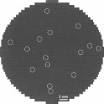
For pharmaceutical applications, imaging Raman spectroscopy enables a spatial analysis of the active and auxiliary substances in newly developed solid formulation. In the following example, we examined the compositional distribution in a cross-sectioned flecainide tablet.