


Intraoperative assessment of resection margin status using X-rays has long been standard in breast cancer treatment in countries such as the USA, China, Japan, and Great Britain [1] [2]. Although the procedure can significantly reduce the number of repeat operations, the technology has been adopted only hesitantly in Germany [3]. This is surprising, given that a reduction in reoperations not only reduces costs but also improves the quality of life for the affected patients.
Is the cut edge free of microcalcification?
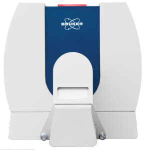
A typical feature of ductal in situ carcinoma is the presence of microcalcifications. These microcalcifications are clearly visible on X-ray images. In breast cancer treatment, the marked tissue is removed during surgery. A compact X-ray machine allows for direct and rapid examination of the excised tissue margin. Once it is confirmed that the excised margin is free of microcalcifications, the wound is sutured and the surgery is completed.
Dr. Rachel Jendro, Lincoln Surgical Hospital, Nebraska, USA
Expert talks about intraoperative margin assessment
Are you interested in specimen radiography?
Sources
[1] Wanheng Li, Xiru Li (2022): Development of Intraoperative Assessment of Margins in Breast Conserving Surgery: a Narrative Review, in Gland Surgery Volume 11, Issue 1, p258-269.
[2] Chantal Reyna, Sarah M. DeSnyder (2018): Intraoperative Margin Assessment in Breast Cancer Management, in Journal Surgical Oncology Clinics, Volume 27, Issue 1, p155-165.
[3] The possibility of intraoperative assessment of the surgical margin using X-rays is not mentioned by relevant German-speaking professional societies:
[3.1] Guideline Program Oncology, S3 guideline for breast cancer, Version 4.3, June 2021.
[3.2] German Cancer Research Center Heidelberg, Cancer Information Service, Surgery for breast cancer, 24.01.2022.
[3.3] German Cancer Society & DIGIMED Verlag GmbH, Onco-Internet portal, Breast cancer surgery, 20.07.2022.