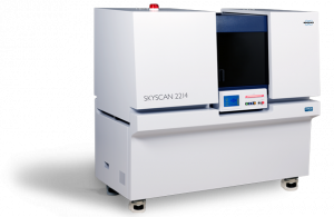
New Nano-CT SkyScan 2214 successfully put into operation
As part of the DELFIN joint project, we have acquired a new type of X-ray microscope (“nano-CT”) to examine the electrode structure of a modern lithium solid-state battery using 3D imaging.


As part of the DELFIN joint project, we have acquired a new type of X-ray microscope (“nano-CT”) to examine the electrode structure of a modern lithium solid-state battery using 3D imaging.

In addition to our daily analysis work, researching and developing new processes is particularly exciting for us. We are therefore very pleased that we are able to work on the DELFIN joint project and thus on an interesting topic - the development of solid-state batteries.
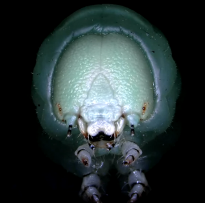
Our customer Dr. Anton Windfelder from the Justus Liebig University of Giessen has published an impressive study on the intestinal tract of the tobacco hornworm caterpillar (Manduca sexta). This animal serves as a model system for ecotoxicology, immunology and intestinal physiology.
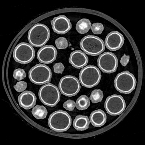
In the Bruker AXS webinar series, our managing director Dr.-Ing. Markus J. Heneka spoke as an expert about the application of 3D X-ray tomography in the pharmaceutical industry. The event was viewed very positively by well over 100 registered participants from all over the world.
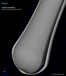
A particular challenge is the correct analysis of microparticles inside medical glass ampoules. When the vials are opened, glass breakage often occurs, which can get into the analysis and falsify the result. To solve the problem, our team has developed a new process to non-destructively detect foreign particles in glass ampoules.
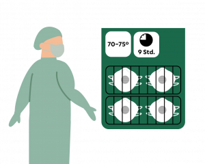
Given the continuing shortage of protection materials for hospital staff during the Corona crisis, Helios experts have in the past weeks developed a safe procedure for refurbishing FFP masks. Helios is making this method publically available, so that all medical staff can be supplied with sufficient protective materials.

Our customer Dr. Arnold Staniczek and his team from the State Museum of Natural History in Stuttgart recently achieved a scientific sensation.
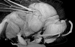
A look inside an insect skull – what zoologists used to only be able to do with a histological examination can now be easily realized using microtomography with multiple magnification and a 3D view. We examined the sample shown here in a SkyScan 1272 micro-CT with a resolution of 1 µm/voxel.