New Nano-CT SkyScan 2214 successfully put into operation
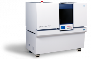
As part of the DELFIN joint project, we have acquired a new type of X-ray microscope (“nano-CT”) to examine the electrode structure of a modern lithium solid-state battery using 3D imaging.
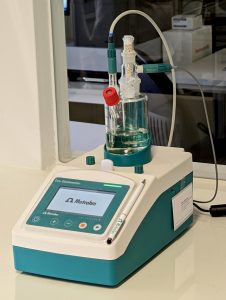
Customers have repeatedly asked us whether we can determine the residual moisture in coolers. We have now equipped our laboratory with a device for analyzing residual moisture and offer this test as standard. We have already successfully carried out the first tests on behalf of customers
In addition to our daily analysis work, researching and developing new processes is particularly exciting for us. We are therefore very pleased that we are able to work on the DELFIN joint project and thus on an interesting topic - the development of solid-state batteries.
Our customer Dr. Anton Windfelder from the Justus Liebig University of Giessen has published an impressive study on the intestinal tract of the tobacco hornworm caterpillar (Manduca sexta). This animal serves as a model system for ecotoxicology, immunology and intestinal physiology.
Our particle scanner for quick and easy cleanliness checks according to VDA-19.1 and ISO-16232 has received an update. The new hardware is even more compact and delivers razor-sharp images of unwanted residual dirt particles with a resolution of 5 µm/pixel.
In the Bruker AXS webinar series, our managing director Dr.-Ing. Markus J. Heneka spoke as an expert about the application of 3D X-ray tomography in the pharmaceutical industry. The event was viewed very positively by well over 100 registered participants from all over the world.
A particular challenge is the correct analysis of microparticles inside medical glass ampoules. When the vials are opened, glass breakage often occurs, which can get into the analysis and falsify the result. To solve the problem, our team has developed a new process to non-destructively detect foreign particles in glass ampoules.
Coxem has introduced a new model of its compact ion mill CP-8000+. The device supports the standard cutting (“cross-sectioning”) and polishing (“flat milling”) modalities and can be operated intuitively via the integrated touchscreen.
A year and a half ago we installed a Coxem EM-30N at the Eberswalde University for Sustainable Development (HNEE). This mosaic shows a 3-point bending fracture in a deformed wood sample. With the standard integrated panorama module from Coxem, 169 individual images of a microtome section of the fracture site were collected at 200x magnification with the secondary electron detector and automatically stitched.