AI segmentation expands the scope of automated SEM-EDX analytics
From
Sebastian Wex
The goal of automated particle and inclusion analysis using SEM-EDX is to detect and examine many objects as quickly as possible (see Phenom ParticleX (from Thermo Fisher). The initial step of object recognition is crucial for the success of automation.
Classical segmentation
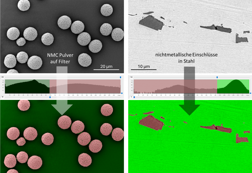
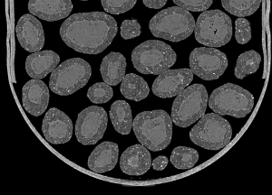
Traditional object detection relies on a grayscale threshold. The BE image is ideal because it shows the material contrast. This allows inorganic particles on an organic filter material to be easily detected using a "lighter than" threshold, and oxide inclusions in metals using a "darker than" threshold.
Analysis of battery powder and steel inclusions using classical segmentation
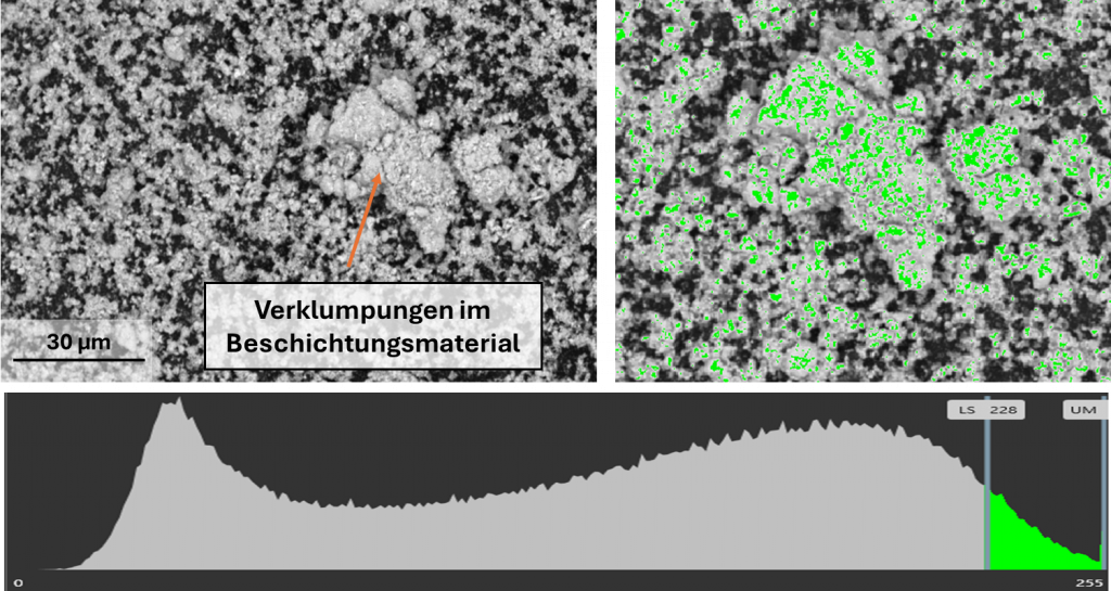
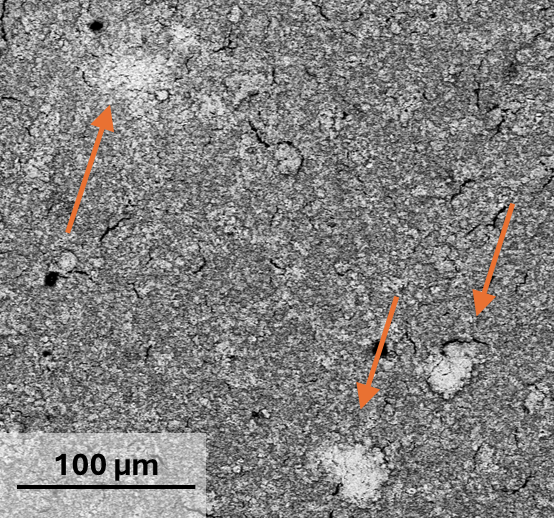
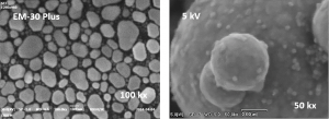
This approach reaches its limits when the objects differ only slightly from the background. The following sample is a platinum-coated foil. The aim of the analysis was to detect areas with clumped platinum islands, as these negatively affect the subsequent process. The clumps are easily recognizable to the human eye in the image. However, automated detection via the grayscale is not possible because the surrounding material is also platinum.
Segmentation using grayscale does not detect the clumping in the coating.
Segmentation supported by AI
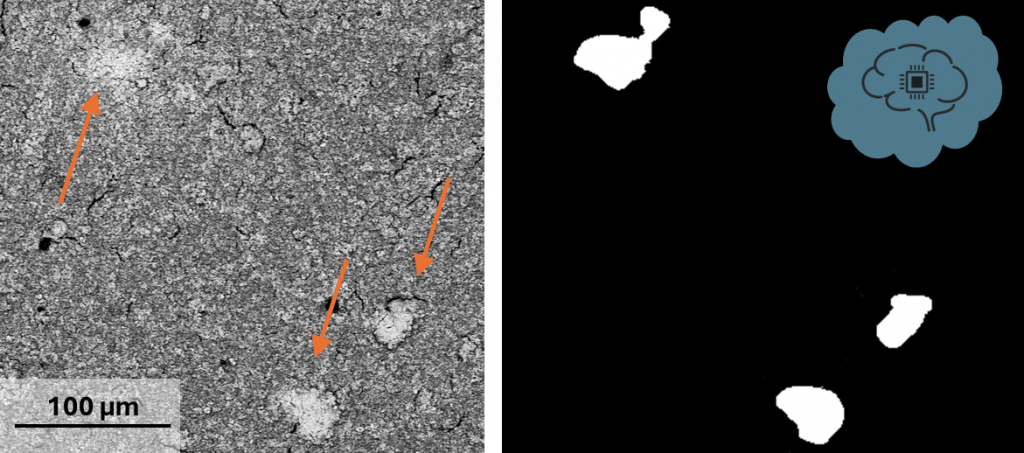
For the Phenom ParticleX Thermo Fisher recently developed an AI-powered segmentation technology that enables object recognition without clear grayscale separation. To use this method, the user must first train a custom-configured AI model using 30 to 40 individual images. The model is then able to differentiate the clumps from the background. This allows for fully automated defect detection and measurement of the number, size, shape, and chemical composition of the defects.
An individually trained AI model specifically detects the clumped zones in the coating.
Another advantage in this specific task is the large sample chamber of the Phenom ParticleX SEM-EDX. This made it possible to analyze the customer's entire 100 x 100 mm sample without cutting it and to statistically record the clumping over a very large area. Here are the quantitative results: