Microtomography enables the examination of Antarctic fish on the Polarstern expedition
From
Markus J. Heneka
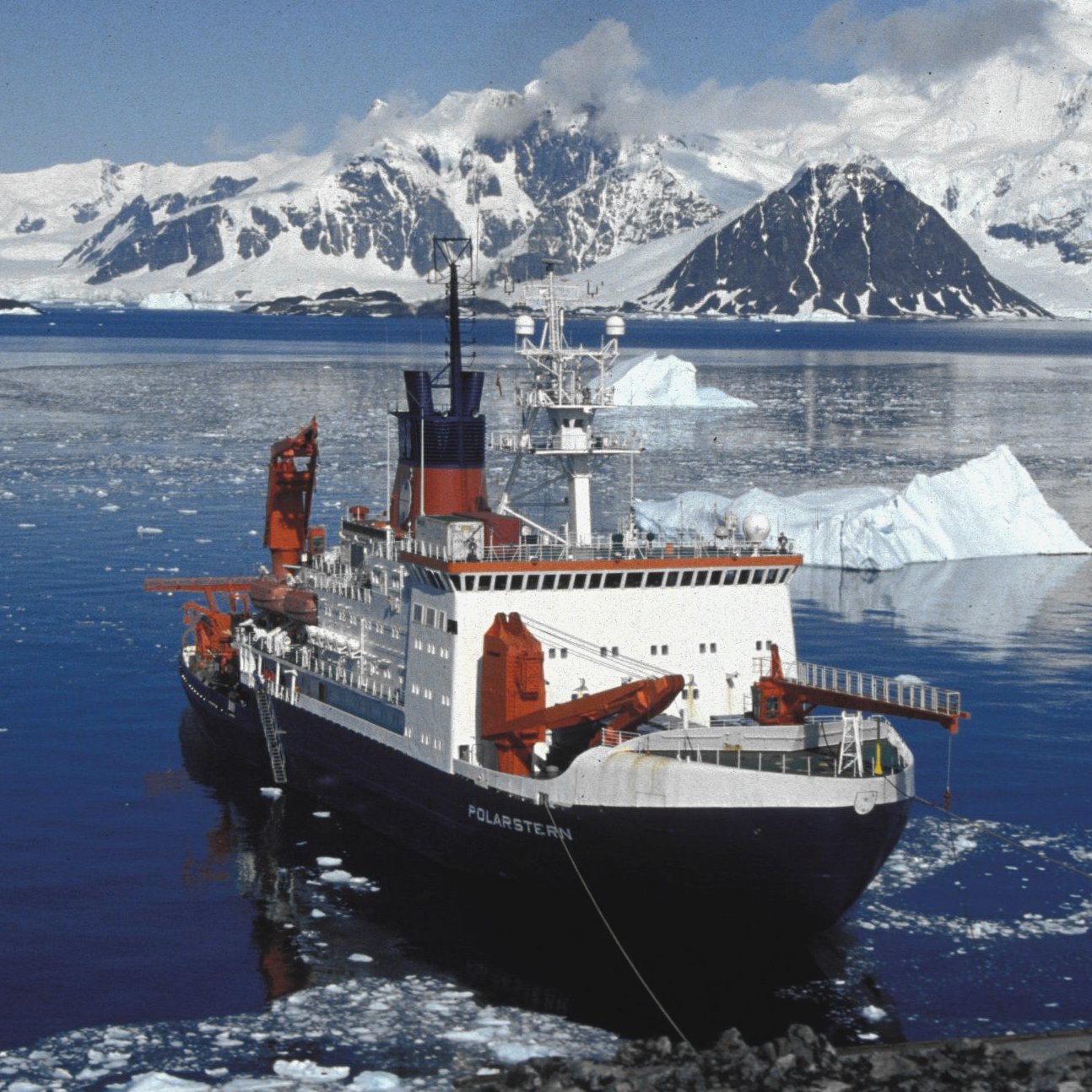
A Bruker micro-CT scanner found a particularly useful application during the Polarstern expedition in the winter of 2025/2026. Christian Bock and his team from the Alfred Wegener Institute Helmholtz Centre for Polar and Marine Research took the micro-CT scanner to the Arctic Ocean to examine the anatomy of Antarctic organisms that are too fragile to be transported. The fact that such a precise mechanical instrument could deliver accurate image data in the rough Antarctic waters was by no means certain beforehand and was only demonstrated during the course of the expedition.
Christian Bock on the research trip, iceberg on the horizon
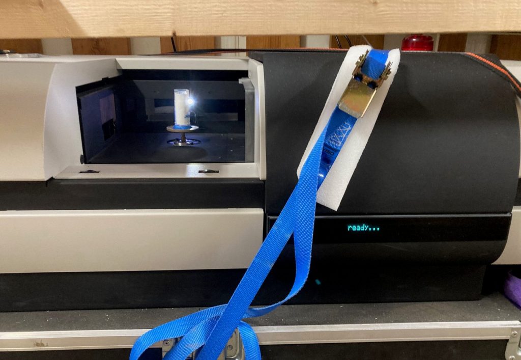
In the calm waters of the Bremerhaven dock, our technicians loaded the micro-CT scanner onto the research vessel in November 2025 and put it into operation. To secure the instrument against the strong waves during the voyage, we lashed it firmly to the ship's hull with ratchet straps.
Bruker micro-CT scanner permanently moored to the Polarstern
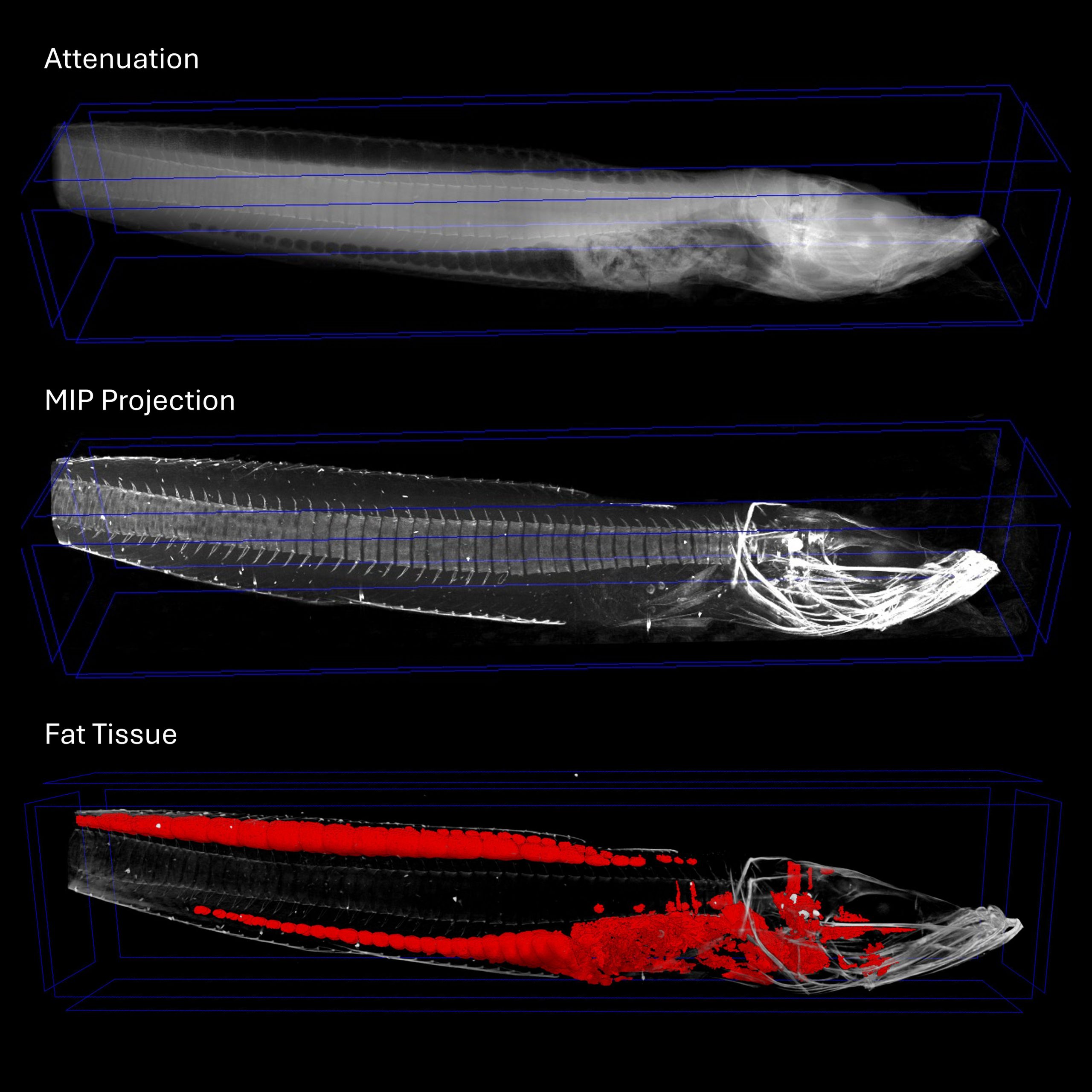
Among other things, the Antarctic silverfish, Pleuragramma antarcticum, measured using micro-CT (see figure). This is a fish species specially adapted to polar conditions and found only in Antarctica. It lacks a swim bladder but has a very high fat content and can survive in water temperatures below zero degrees Celsius. Due to its high fat content, it is an important food source for seals and penguins. Christian Bock collected several specimens from various depths of up to 800 meters in the Weddell Sea. The animals were then scanned using micro-CT to examine the fat distribution. The following figure shows a clear example of this analysis:
Micro-CT scan of an Antarctic silverfish, fatty tissue in red
We are pleased that the measuring setup functioned so reliably under the stormy conditions and would like to thank Christian Bock for providing the image material and the interesting insights into his work.