


The discovery of a mummy or unique artifacts of human history is a sensation because it forms part of the cultural heritage. For many investigations, however, a damage of the object, for example in the form of a sampling is required, the preservation of the find for science is no longer guaranteed. Archaeologists and representatives of related disciplines have been using X-ray microtomography for several years to provide a method that allows them to be examined non-destructively.
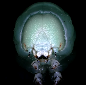
With this method, scrolls can be unrolled virtually, with charred woods, the wood species can be determined. The observation of plant remains and insects with the micro-CT, however, provides information about their taxonomy. Researchers also have tools, pottery shards, other grave goods or teeth analyzed by micro-CT. On bones such as deformations, injuries or even diseases can be determined.
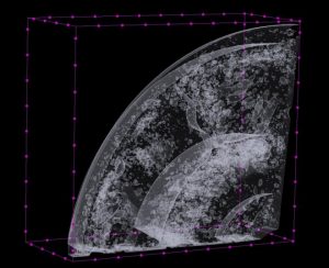
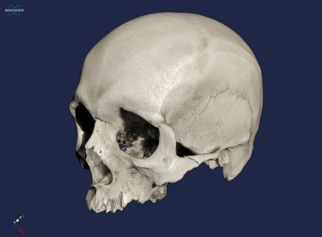
The following video shows the scan of the skull of a full-grown male individual from Thuringia, dated to the later Middle Ages, ca. 13th century. The sample was provided to us by employees of the University of Jena. The skull is about 20 x 20 cm tall, the object was examined with the Multiscale Nano-CT SkyScan 2211.
Find out about the SkyScan devices from Bruker microCT and about our accredited analysis service.