Osseointegration of metallic implants depicted without artifacts
From
Markus J. Heneka
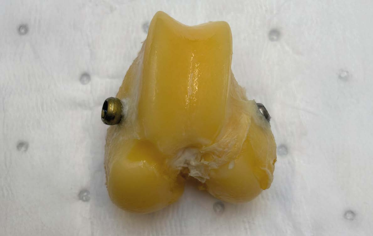
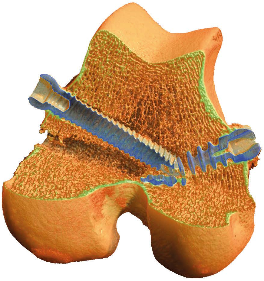
Various implants, including titanium or steel screws, can be used in the surgical treatment of bone fractures. The screw fixes the fragmented bone. Experiments on animal bones can be used to investigate the performance of the implants and their integration into the bone. This requires an examination procedure with optimal imaging. The new X4 POSEIDON X-ray micro-CT scanner from Bruker offers the best conditions for this. Our test subject is a distal femur from a sheep with two titanium screws.
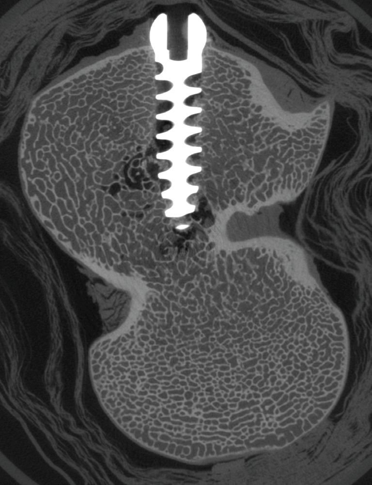
We scanned our specimen with the X4 POSEIDON at an X-ray energy of 110 kV, a 1 mm molybdenum filter, and a voxel size of 30 µm. The cross-sectional image through a titanium screw in the femoral condyle shows no artifacts in the shadow of the metal. The undamaged bone structure in the screw thread is clearly visible.
In orthopedic research, the X4 POSEODON Desktop Micro-CT platform offers ideal conditions for investigating the function and performance of bone implants. Would you like to learn more? Please contact us!