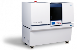


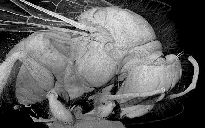
A look into the inside of an insect's skull - what used to be possible only for zoologists by means of a histological examination can be conveniently realized on the basis of microtomography with multiple magnification and a 3D view. We have the sample presented here in a Micro-CT of the type SkyScan 1272 examined with a resolution of 1 μm / voxel and visualized the internal incision of the masticatory muscles.
It is the head of a mayfly nymph of the genus Coloburiscus, whom employees of a German natural history museum have sent to us for examination. For the determination of an object, the zoologists create a taxonomy by classifying the animal according to certain criteria and thus determining similarities and distinguishing features. In zoology, the method of microtomography has been proven in recent years as a method for the morphological characterization of animal and vegetable samples.
Find out about the SkyScan devices from Bruker microCT and about our accredited analysis service.